Back to productS

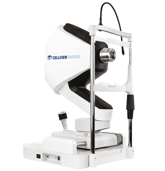
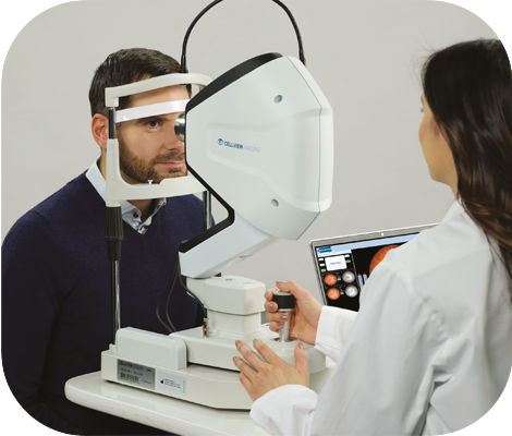
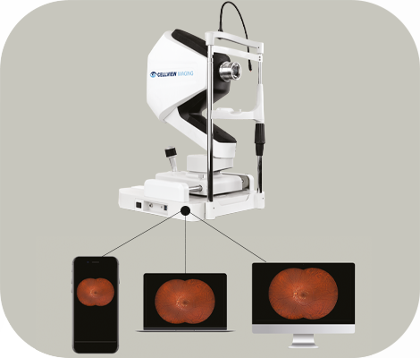
WRI-1 Widefield Non-Mydriatic
Retinal Imager


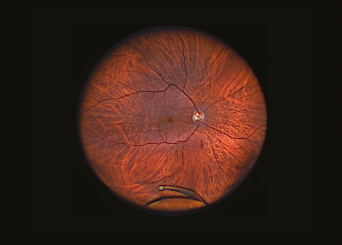
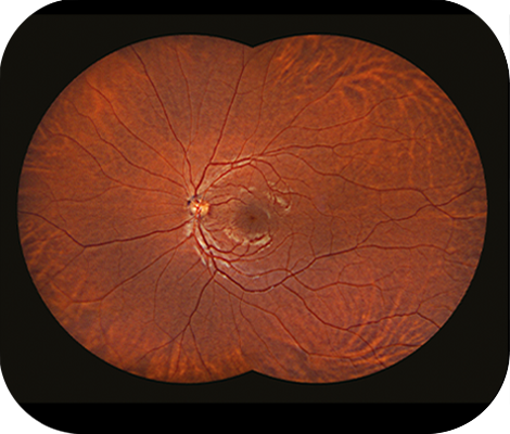
The WRI-1 captures a retinal image and far into the periphery up to 133° in a single-capture, or an up to 200° auto-stitched image.

It provides clear high defination¹ images, enabling examiners to image patients who are traditionally challenging, such as those with small pupils, cataracts or other media opacities.

Download brochure

 Why WRI-1?

High-definition widefield fundus imaging

Screen retinal defects or abnormalities at a stage when intervention and treatment are most effective.

The WRI-1 features a patient friendly, Low-intensity illumination system that enables imaging through pupils as small as 2.5mm without dilation.

The device has the ability of taking Full Color and Infrared retinal imaging.

Screening and evaluation of vision-threatening conditions

The full visible spectrum LED array provides both full colour and infrared images for screening potential retinal abnormalities.

  • Easy-to-operate device with an intuitive user interface.
  • Patient-friendly capture for smoother clinic workflow.

Automatically adjusts focus and gain for sharp retinal images streamlining the imaging process.

Extensive Cloud Storage & Remote Review Acess

The extensive cloud storage solution, Remote Review Station and Web Review, make images accessible whenever and wherever you need it. 

Minimize downtime and costs thanks to remote support when needed.

Stay at the forefront of technology with the latest tools and instant seamless updates.

Disclaimers:
1. Please refer to the Pixel pitch resolution line in the Technical specifications table. Dislocated IOL Retinal tear in periphery and laser surgery.
2. Encrypted data transfer & storage.
3. Feature available according to markets, please refer to your local sales team.